
The Role of the Paleocerebellum in Eliminating Violence in Mother-deprived Primates and Permitting Expression of Affectional Behaviors Not Possible Before Paleocerebellar Surgery.
James W. Prescott, Ph.D.
It is well recognized that Violence by Homo Sapiens throughout the World threatens species and planetary survival. Violence begins with the individual and must be understood before Cultures of Violence appear. The brain is the organ of behavior and how the brain is encoded for Peaceful or Violent Behaviors is the great challenge to Humanity.
It is also well recognized that Pain and Pleasure experienced during the formative periods of brain development determine the life path of the mammalian organism that is carried throughout life. How Pain and Pleasure are encoded in our two developing brains: 1) subcortical emotional-social sexual brain, first in evolution and development; and 2) the neocortical rational, thinking brain that mediate our values, which is second in evolution and development, determines the life path of Peace or Violence that will be followed.
These evolutionary-developmental processes are governed by the principle of reciprocal inhibition that governs all behavior.
An organism in a state of Pain is not in a state of Pleasure. An organism in a state of Pleasure is not in a state of Pain. If an individual is Happy, he/she is not depressed. An angry person inhibits a joyful person. An egalitarian person inhibits the authoritarian person. A peaceful person inhibits the Violent person. Pain is a Moral good. Pleasure is a Moral evil.
Aristotle (c384-322 B.C).stated in Nichomachean Ethics, Book 7:
“Therefore, the highest good is some sort of pleasure, despite the fact that most pleasures are bad, and, if you like, bad in the unqualified sense of the word”
These relationships are presented in Our Two Cultural Brains
http://www.violence.de/prescott/letters/Our_Two_Cultural_Brains.pdf and at:
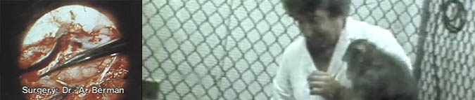
The significance of this paleocerebellar decortication is not only found in the elimination of violent pathological behavior in the mother deprived adult primate but equally important is the expressing of affectional relationships, as evidenced in the touching and hand-feeding of this primate, behaviors not possible before surgery.
Of particular importance are the hand-feeding and climbing on the back of the technician, which is aberrant social behavior that is not observed in the normal reared feral primate. This primate never engaged in this behavior before in its life nor observed this behavior in other primates. Where did this “social-learning” come from? Certainly not from its genes!
The cerebellar study published by Berman, Berman and Prescott (1974)
Berman, A.J., Berman, D. & Prescott, J.W. (1974). The effect of cerebellar lesions on emotional behavior in the rhesus monkey. In: The Cerebellum, Epilepsy and Behavior. (Cooper, I.S., Riklon, M.V. & Snider, R.S. (Eds) Plenum, NY
http://www.violence.de/berman/article.html
and the cerebellar Pacemaker studies by Heath, et al. to control violence in untreatable psychiatiric patients require an “agonizing reappraisal of this research and findings by the National Institutes of Health.
Robert G. Heath, D.Sc., M.D.: Gross Pathology of the Cerebellum in Patients Diagnosed and Treated as Functional Psychiatric Disorders
The Journal of Nervous and Mental Disease, Volume 167, Number 10 (October 1979), pp. 585-591. PDF format of a 1-bit scan, low quality of tomography reproduction (last modified 2003/11/11).
Heath, Llewellyn and Rouchell: “The Cerebellar Pacemaker for Intractable Behavioral Disorders and Epilepsy: Follow-Up Report”
Biological Psychiatry, Volume 15, Number 2 (1980), pp. 243-256.
(…) The patients who have responded best to the treatment are those with depression, those with behavioral pathology consequent to epilepsy, and those with psychotic behavior consequent to structural brain damage. (…) Twenty-one percent of the patient group displayed structural evidence of cerebellar pathology that was not detected before operation, a finding which suggests that cerebellar damage may induce psychotic behavior. (last modified )
Robert G. Heath, D.Sc., M.D. et al.: Cerebellar Vermal Atrophy in Psychiatric Patients Biological Psychiatry, Volume 17, Number 5 (1982), pp. 569-583.
The following study of Heath (1975) published in the proceedings of a Conference sponsored by the National Institute of Child Health and Human Development (NICHD) deserves special attention, as it documents impaired brain function In mother-deprived primates. What are the implications of these findings for homo sapiens?
Heath, R. G. (l975): Maternal-social deprivation and abnormal brain development: Disorders of emotional and social behavior. In Brain Function and Malnutrition: :Neuropsychological Methods of Assessment (Prescott, J.W., Read, M.S., & Coursin, D.B., Eds). John Wiley New York. http://www.violence.de/heath/bfm/article.html
Dr. Collins, given the extraordinary significance of the above studies for the understanding and prevention of violence by Homo sapiens, I am requesting information on the percent of NIH funding on violence research, particularly, the role of cerebellar regulatory mechanisms of emotional behaviors by the various National Institutes of Health.
You should be aware of the report in April 1994: Publication of the “Report of the Panel on NIH Research on Antisocial, Aggressive, and Violence-Related Behaviors April 1994: Publication of the “Report of the Panel on NIH Research on Antisocial, Aggressive, and Violence-Related Behaviors and their Consequences”, that
e) Summary September 22-24, 1993 meeting: 2nd paragraph:
“To date, investment across all Institutes and ICDs in violence-related research has been minuscule relative to the total NIH budget (i.e.0.5%).” View Page 138.
http://www.violence.de/history/NIHR_1994.html
An update of this statistic is requested. What is the current level of NIH support for violence research?
April Dworetz in an essay in the New York Times on End of Life, at Birth documented the high risk environment of the ICU for prematures, stated: For all the biomedical advances, though, the key ethical problems surrounding premature birth remain, which gives added importance to my proposal for rocking bassinetts in the ICU for restoring the maternal-infant/child affectional bond.
http://www.nytimes.com/2013/08/05/opinion/end-of-life-at-birth.html?ref=opinion
Jan Hoffman, wrote in a New York Times essay,: Nightmares After the ICU, July 22, 2013 of the Sensory Deprivation of the I.C.U and the hallucinations (PTSD) such sensory deprivation induces:
When Lygia Dunsworth was sedated, intubated and strapped down in the intensive care unit at a Fort Worth hospital, she was racked by paranoid hallucinations:
Outside her window, she saw helicopters evacuating patients from an impending tornado, leaving her behind. Nurses plotted to toss her into rough lake waters. She hallucinated an escape from the I.C.U. — she ducked into a food freezer, only to find herself surrounded by body parts.
http://well.blogs.nytimes.com/2013/07/22/nightmares-after-the-i-c-u/?pagewanted=print&_r=0
Sensory deprivation of immobilization has been well described in the psychological research literature that induces hallucinations and depersonalization:
IMMOBILIZATION—SENSORY DEPRIVATION
and Emotional Behavioral Disorders
It would be remiss of me not to mention in this section on immobilization the findings of Zubeck (1969) and Zubeck, J.P., Aftanas, M, Kovach, K., Wilgosh, L..). and Winokur, G. (1963) who immobilized adult faculty subjects for periods of up to 24 hours (mean – 12.9 hours; S.D. -6.6. hours). Only 8 of their 40 subjects were able to endure immobilization for the prescribed 24 hours. Vision and hearing was not interfered with and social interaction with the experimenters was maintained. Head movements were prevented with a head-holder. These investigators reported that 85% found immobilization stressful and that 75% stated they would not repeat the experience in a week’s time. The severe restrictions of kinesthetic (proprioceptive and vestibular) activity alone resulted in there showing “intellectual inefficiency, bizarre thoughts, exaggerated emotional reactions, time distortions, changes in body image, unusual bodily sensations and various physical discomforts, than the recumbent controls” (p.127). Specifically, subjects felt “that some part of their body was disconnected or did not belong to the rest of the body, that they were melting or merging or merging into their surroundings, and that at times they felt like a different person,” (p.126). Reports included “whole body floating or revolving in space, whole body feels as heavy as a “ton of bricks” and various distortions of body properties such as one limb being much shorter than another” (p.126). Psychosomatic complaints were also frequent and included periodic aches and pains, numbness, dizziness, physical discomfort, chills, perspiration, weakness; strong desire to scratch parts of the body and difficulty in sleeping. These physical symptoms were responsible for almost all of the early terminations of the experiments prior to the prescribed 24 hours.”(pp. 126-127). Additional data are summarized elsewhere( Appley, 1967; Zubeck, (1969).
It is apparent from the above that severe somatosensory deprivation in healthy adults for less than 13 continuous hours has profound effects upon the integrity of intellective, emotional and somatic functions. Disintegration of integrative functioning would seem to aptly describe the above effects of somatosensory deprivation. It would seem appropriate to inquire as to the effects of “insufficient” somatosensory stimulation during critical periods of development, particularly upon maturation of integrative functioning. What constitutes “sufficient” or “insufficient” somatosensory deprivation for the developing infant appears to be an issue of extraordinary importance and major research efforts should be directed to that question. (Prescott, 1971, 1976, pp. 74-75)…
The symptoms reported above are remarkably similar to those reported by Hoffman (2013) in the I.C.U.
References
Bell, C. and Dow, R.S..Cerebellsr Circuitry. Neuroscience Research, Program Bulletin 5 (2): 121-122.
Anand, B.K. ,Malhotra, D.L, Xingh, B. and Dua, S. (1959). Cerebellsr projectins to limbic system. J, Neurophysiology. 22 (4): 451-457.
Prescott, J.W. (1976). Somatosensory deprivation and its relationship to the blind. In: The Effects of Blindness and Other Impairments on Early Development pp.65- 121(Z.S. Jastrembke, Ed.). American Foundation For The Blind, New York.
http://www.violence.de/prescott/afb/paper.pdf
Zubeck, J. P., Aftanas, M, Kovach, K., Wilgosh, L.. and Winokur, G. (1963) Effect of severe immobilizsation on intellectual and perceptual processes. Canadian Journal of Psychology. 17:118-133.
Zubeckj, J.P. (Ed). (1969. Sensory Deprivation: Fifteen years of Research. Appleton-Century Crofts, New York.
Institutionalization as Sensory Deprivation.
Dennis (1973) describes the profound deprivation of sensory stimulation in early development, Children of the Creche. He noted “babies from birth to about one year spent most of their time in bassinets. During most of this year they were fed by bottle in their cribs; and were almost never taken from their cribs except for daily bathing and change of clothing. (Prescott, 1976, P.76)
Not much different than the modern ICU for Prematures.
Pediatric Immobilization, Sensory Deprivation and Emotional Disorders.
Additional clinical data on the effects of somatosensory deprivation on children have been provided by Freedman, et al (1968) and Freedman (1968). They demonstrated that immobilization of infants and children with plaster casts; traction, Dennis-Brown splints and other restraint procedures resulted in emotional disturbances of depression mixed with hyperactivity and outbursts of violence. Sibinga and Friedman (1971) also reported delays in language development in children with a history of immobilization (also reports of impaired pain perception), as noted in maternally deprived monkeys, Sacket (1970; Mason and Berkson, 1975).
Sibinga has also observed gastric ulcerations in children who have been extensively immobilized subsequent to severe burn injuries or CNS surgery and that a lesser degree of immobilization frequently resulted in chronic idiopathic diarrhea…In this context, the findings of Wolfe (1969) that lesions in the cat cerebellum produces gastric ulceration is notable. (Prescott, 1976, p.71).
Dr, Collins, you are requested that the information provided to you in this BLOG and in the New York Tmes be reviewed by you and the Institute Directors concerned with the ICU environment and the treatment of prematures with a statement of health policy changes that this information dictates.
References
Appley, M.H. and Trumbull, R. (Eds) (1967). Psychological Stress. . Appleton-Century Crofts. New York.
Dennis, W. (1973). Children of the Creche. Appleton-Century Crofts. New York.
Freedman, D.A.. (1968). The Influences of congenital and perinatal sensory deprivation on later development. Psychosomatics. 9 (5: 273-277.
Friedman, C.J., Sibinga, M.S., Steisel, I.M. and Sinnamon, H.M. Sensory restriction and isolation experiences in children with phenylketonuria. J. Abnormal Psychology. 73(4):294.303.
Mason, W.A. and Berkson, G. (1975). Effects of Maternal Mobility on the Development of Rocking and Other Behaviors in Rhesus Monkeys: A Study with Artificial Mothers. Developmental Psychobiology. 8, 197-221
http://www.violence.de/mason/mason74.pdf
Sibinga, M.S. and Friedman, C.J. (1871) Restraint and speech. Pediatrics 48 (1): 116-122.
Sackett, G.P. (1970) Unlearned responses, differential rearing experiences and the development of social attachments by rhesus monkeys. In. Primate behavior.: Developments in field and laboratory research. Vol !. (Rosenblum, L.., Ed.) Academic Press. New York.
Wolfe, J.W. (1969). Chronic gastric ulcers associated with experimentally induced posterior cerebellar vermal lesions. Physiology and Behavior 4: 1011-1019.
Figure 1 is a photo collage of the devastation of child abuse inherent in maternal-infant/child separation.
I am copying this BLOG to the New York Times Science writers for their interest in bringing this information to the public.
Collins letter OF 11 July 2013 HERE
BIOBEHAVIORAL SYSTEMS
1140-23 Savannah Road
Lewes, DE 19958
302.645.7436
jprescott34@comcast.net
http://www.violence.de
http://montagunocircpetition.org
11 July 2013
Francis S. Collins, M.D., Ph.D.
Director, NIH
National Institutes of Health
9000 Rockville Pike
Bethesda, MD 20892
Dear Dr.Collins,
This letter is a follow-up of my letter to you of 6 June 2013 that requested a review of my letter to Jeffrey M. Drazen, M.D. Editor-in-Chief New England Journal of Medicine with a copy of my DVD: The Origins of Love &Violence: Sensory Deprivation and the Developing Brain with a request that you install rocking bassinets in all ICUs in this country, which would compensate for vestibular-cerebellsr sensory deprivation that prematures experience in utero and post-natal development resulting in increased neural maturation and survival. I have not received a reply from you concerning this letter.
The scientific foundations for the above research and recommendations are to be found in the published works of Cannon (1939); Cannon and Rosenbleuth (1949) and Sharpless (1969, 1975), which, unfortunately, are relatively unknown in the biomedical science communities.
Of particular relevance here are the findings of Berman, A.J., Berman, D. & Prescott, J.W. (1974). The effect of cerebellar lesions on emotional behavior in the rhesus monkey. In: The Cerebellum, Epilepsy and Behavior. (Cooper, I.S., Riklon, M.V. & Snider, R.S. (Eds) Plenum, NY http://www.violence.de/berman/article.html
And in the video documentation of this study:
Prescott, J.W. (1976). Cerebellar surgery: abolishment of pathological violence in the adult rhesus mother= deprived monkey with petting and hand-feeding not possible before surgery. W- 5, CTV Toronto. November 11 minute film clip War on Women and Children.
The continuing and unconscionable prematurity and infant mortality rate of this nation calls for drastic action of leadership that appears lacking in the biomedical communities.
I am copying this letter to the respective and relevant NH Institute Directors and to
Jeffrey M. Drazen, M.D. Editor-in-Chief New England Journal of Medicine and nejmcust@mms.org
Lewis R. First, M.D.
Editor-in-Chief, PEDIATRICS
lewis.first@uvm.edu
It is requested that you distribute this email and Blog to those that should be knowledgeable of these issues and to encourage Drs Drazen and First to to alert their membership of this BLOG.
Sincerely,
James W. Prescott, Ph.D.
Director.
For those not familiar with my research activities on behalf of women and children, I am attaching a brief BIO of that work.
Prescott, J.W. (1977). Phylogenetic and ontogenetic aspects of human affectional development. In: Progress In Sexology. Proceedings of the 1976 International Congress of Sexology. (R. Gemme & C.C. Wheeler, Eds.) Plenum Press, New
York http://www.violence.de/prescott/pis/1977paper.pdf